
Foot drop
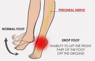
Foot drop, which is also sometimes called a dropped foot, is a condition in which it is difficult to lift up the front part of your foot. As a result your forefoot and toes tend to catch or drag on the floor as you walk. It can be temporary or permanent and most commonly affects one side only.
The gait of foot drop may involve:
- As you walk along, the affected foot (or feet) catches on the floor.
- As you walk along you lift the leg high to avoid the foot catching (high stepping gait). People who do this often tend to walk on tiptoe on the other side to equalise the sides.
- As you walk you swing the affected leg out to the side to avoid it catching on the floor.
Foot drop usually affects just one foot. However, it can affect both sides, either equally or to different degrees. It may be temporary or permanent.
There are several grades of foot drop. These are measured from 0 to 5 depending on the degree of strength and movement there is in the muscles which lift the foot. 5 is normal strength and 0 is total paralysis.
Causes
Foot drop is usually caused by malfunction of a nerve in the lower leg. This can be due to problems affecting it either low down in the leg, or higher up in the spine where its nerve fibres originate.
This nerve is called the common peroneal nerve. However, it is also sometimes called the common fibular nerve, the external popliteal nerve or the lateral popliteal nerve. It’s a small nerve that branches off from the sciatic nerve in the thigh. It runs down the back of the knee and winds around the top of the fibula to go into the muscles of the lower leg. It is very near to the surface at this point and can be easily bruised or compressed.
The most common causes are:
- Injury to the common peroneal nerve.
- Lower back damage (including a ‘slipped’ disc (prolapsed disc)affecting the nerves in the lower leg).
Foot drop can also be due to other causes of nerve damage. More rarely, it can be due to damage to the muscles of the lower leg or to poisonous substances or a tumour.
Other causes include:
- Hip replacement.
- Knee surgery.
- Sciatic nerve damage.
- Cauda equina syndrome. (This is compression of the nerves in the tail of the spinal cord, usually caused by a ‘slipped’ disc or tumour.)
- Diabetes with peripheral neuropathy.
- Stroke.
- Transient ischaemic attack (TIA).
- Multiple sclerosis (MS).
- Cerebral palsy.
- Charcot-Marie-Tooth disease.
- Poliomyelitis (rarely causes isolated foot drop).
- Motor neuron disease.
- Friedreich’s ataxia.
- Brain tumour.
- Adverse drug or alcohol reaction.
Symptoms
People who have foot drop may drag their toes when they walk. They may also have to lift their knees higher than usual to avoid dragging their toes. Other symptoms include muscle weakness and “tingling” feelings in the leg.
Diagnosis
Foot drop is usually diagnosed on examination. Your doctor will watch you walk and may check your leg muscles for weakness. They will also assess nerve function by checking your reflexes and the sensation in the skin.
Foot drop is a symptom rather than a diagnosis and your doctor will want to understand what has caused it. Investigations for the cause of foot drop may include:
- X-rays. Plain X-rays may be used to look for a soft tissue growth or a bone abnormality that may be causing your symptoms.
- Ultrasound. This may be used to check for cysts or tumours that may be pressing on the nerve.
- Computerised tomography (CT) scan. This can be used to look for growths (masses) or other changes anywhere in the body.
- Magnetic resonance imaging (MRI). MRI is particularly useful in visualising soft tissue lesions that may be compressing a nerve, such as a ‘slipped’ disc (prolapsed disc) in the back. It can also examine the brain for the characteristic lesions of MS.
- Electromyography (EMG) and nerve conduction studies. These measure electrical activity in the muscles and nerves to look for the location of the damage along the affected nerve.